Prostate matters is a not for profit organisation committed to providing free information about prostate issues from leading Clinical Authorities.

Multi parametric MRI (mpMRI) in prostate cancer diagnosis

Consultant Uro Radiologist
University College Hospital London, NHS Trust
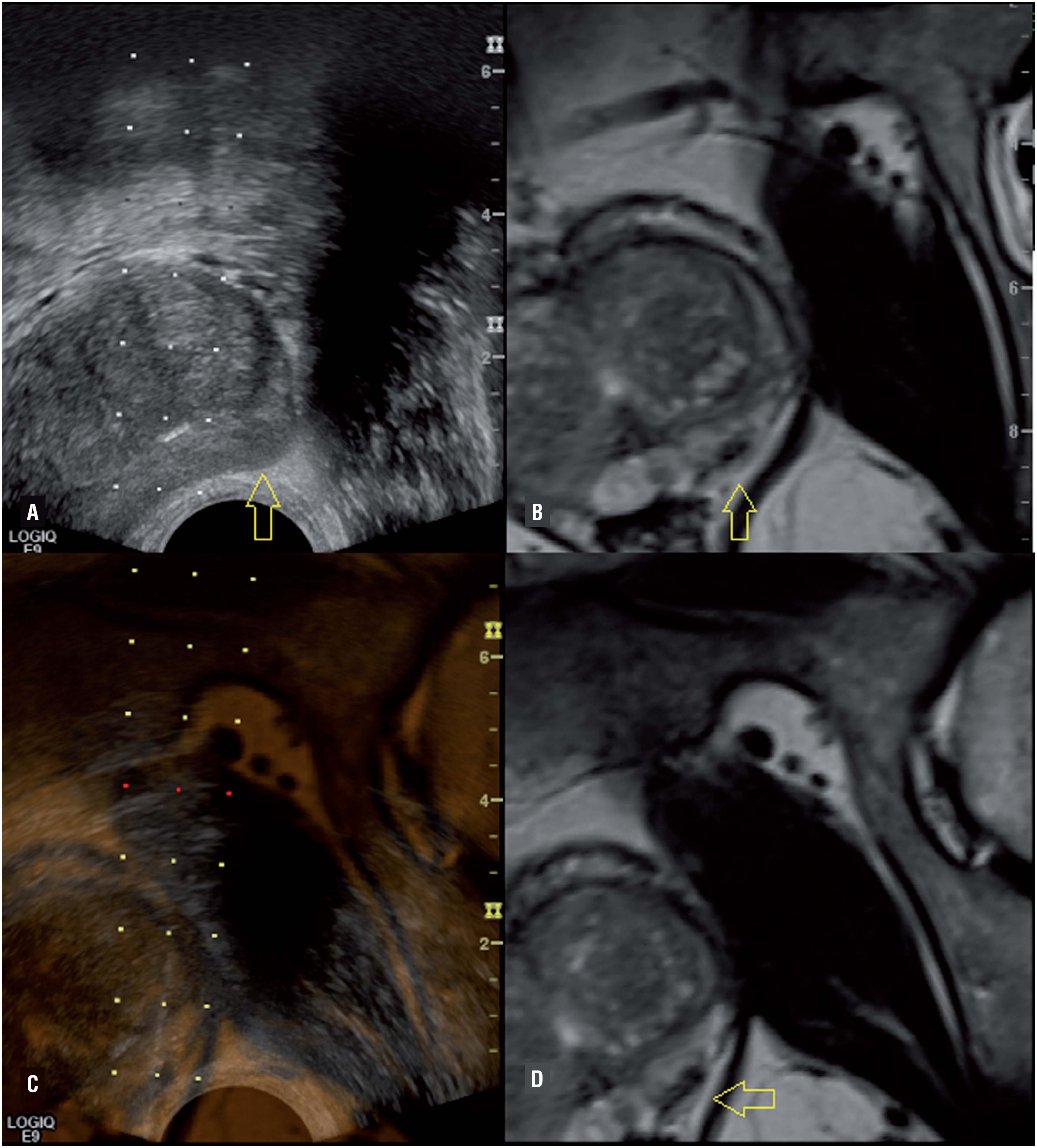
There have been substantial improvements with MRI in Prostate Cancer diagnosis in recent years, the most significant being the introduction of multiparametric MRI scans (mpMRI).
Advances in MRI technology mean that many modern MRI scanners provide satisfactory scan quality. The Consensus Paper (see link) recommends a 3T scanner as being optimal (T stands for Tesla – a measure of magnetic strength. 1T is 10,000x the earth’s magnetic field, so 3T is 30,000 times), although newer 1.5T scanners (which are less powerful) are good enough.
The Consensus Paper has been written jointly by a combination of leading urologists, radiologists, pathologists, and oncologists, with input from radiographers and physicists, to produce a document that lays out the specific parameters around imaging the prostate in the context of suspected or actual prostate cancer.
High quality mpMRI scans enable specialist experienced radiologists to differentiate between benign and potentially malignant conditions with confidence.
mpMRI is now the recommended diagnostic step prior to biopsy. It has several advantages for patient comfort and diagnostic efficacy compared with other techniques.
The PROMIS trial testing the effectiveness of mpMRI
Between May 17, 2012, and November 9, 2015, 740 men were enrolled in a trial to test the effectiveness of mpMRI.
For clinically significant cancer, MP-MRI was more sensitive (93%, 95% CI 88–96%) than TRUS-biopsy (48%, 42–55%; p<0·0001)
In around a third of patients (though this depends on the PSA), the MRI is so convincingly negative, that we can say that there is unlikely to be significant disease in the gland. For some men this is reassurance enough to prevent the need for biopsy.
If a biopsy is required, the abnormal area on MRI can be targeted. In particular, the 25-30% of tumours that occur in the anterior part of the prostate can be reliably detected, which had often missed by TRUS Biopsies.
With patients undergoing Active Surveillance, mpMRI can be used to check that there has not been significant disease progression. Given there is little correlation between PSA level and tumour size, mpMRI is a better option than PSA surveillance.
Clinical Evidence of mpMRI Benefits
- The diagnostic and patient benefits of mpMRI diagnostic scanning prior to any potential biopsy
- The surveillance advantages
mpMRI Now Recommended by NICE
As of December 2018, the National Institute for Clinical Excellence (NICE) has recommended mpMRI scans as the the first line investigation for people with suspected clinically localised prostate cancer

Prostate matters is a not for profit organisation that is committed to providing free expert advice about prostate issues from leading Clinical Authorities
In memory of Riki
PROSTATE MATTERS
FOLLOW US
Copyright Disclaimer: We try to acknowledge copyright as appropriate. If we have used something without acknowledging copyright, this is inadvertent. Please let us know by emailing info@prostatematters.co.uk
Site design and technical development by Webtoys | Intelligent Digital Media